MRI without contrast agents? Yes, with sugar!
Scientists from the German Cancer Research Center (DKFZ), in collaboration with colleagues from Heidelberg University Hospital, have been able to visualize brain cancer using a novel MRI method. They use a simple sugar solution instead of conventional contrast agents, which can have side effects in the body.
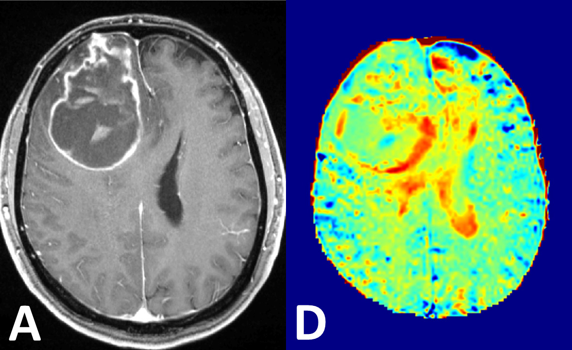
Visualization of brain cancer by conventional contrast agents (left) and by the novel glucose MRI method (right).
© Paech D, et al. T1ρ-weighted dynamic glucose enhanced MRI in the human brain. Radiology (in press). © RSNA
In magnetic resonance imaging (MRI), contrast agents are used to enhance the imaging of tissue structures. While they enhance signals in blood vessels and in spaces between cells, they do not reach the interior of the cell. By contrast, glucose is taken up and then broken down in the body cells. Tumor cells are particularly hungry for glucose in order to feed their high energy needs. By observing glucose metabolism activity it may therefore be possible to identify solid tumors or very aggressively growing tumor areas. Radiologists and physicists from the German Cancer Research Center (Deutsches Krebsforschungszentrum, DKFZ) in Heidelberg have now succeeded in employing this novel type of imaging.
Conventional MRI is based on measuring the signals from protons in water. Since over 60 percent of our body is made up of water, this delivers a clear picture. Glucose is found at much lower levels in our body. In order to make it visible, the DKFZ researchers are therefore using an ultrahigh field scanner with 7 Tesla magnetic field strength and a special method to reinforce the glucose signal distinctly and selectively. This makes it possible to obtain sufficient signal strength in order to visualize changes in glucose levels in brain tissue after injection of a glucose solution.
The underlying physical principle of this method is called magnetization transfer effect. While this effect has been known for decades, it hasn't been possible so far to use it for glucose imaging in humans. In magnetization transfer, the signal from glucose protons is transferred to bodily water, which is measured in MRI. The effect is proportional to the local glucose level, thus reflecting regional changes in glucose levels. The amount of glucose needed for glucose measuring corresponds to about five sugar cubes.
In the present work, physicist Patrick Schuenke and physician and physicist Daniel Paech have been able to observe the changes of glucose signals in healthy brain regions as well as pathogenic changes in human brain cancer.
For decades now, scientists have used another measuring method, called positron emission tomography (PET), to visualize elevated glucose uptake in tumors. However, this method requires radioactively labeled glucose molecules. "Our glucose MRI does not require any radioactivity and therefore does not involve any radiation exposure for the patient," said Paech, who is the first author of the publication.
The glucose MRI project is a collaboration of DKFZ scientists from the groups led by Peter Bachert, Mark Ladd and Heinz-Peter Schlemmer. The researchers have pointed out that some questions about the new measuring method still have to be pursued. "We do not know yet how the shares of measured glucose are distributed between vessels and extracellular spaces on the one hand and the cell interior on the other," said radiologist Heinz-Peter Schlemmer. "If we can confirm that substantial signal levels originate from glucose in the cell interior, this would be important additional information for tumor imaging and functional MRI. This could enhance therapy planning and monitoring."
Paech, Schuenke, Koehler, Windschuh, Mundiyanapurath, Bickelhaupt, Bonekamp, Bäumer, Bachert, Ladd, Bendszus, Wick, Unterberg, Schlemmer, Zaiss, Radbruch. T1ρ-weighted Dynamic Glucose Enhanced MRI in the Human Brain.
Radiology 2017, DOI: 10.1148/radiol.2017162351
Former publications on this topic:
Schuenke, Koehler, Korzowski, Windschuh, Bachert, Ladd, Mundiyanapurath, Paech, Bickelhaupt, Bonekamp, Schlemmer, Radbruch, Zaiss. Adiabatically Prepared Spin-Lock Approach for T1ρ-Based Dynamic Glucose Enhanced MRI at Ultrahigh Fields. Magnetic Resonance in Medicine, 2016, DOI:10.1002/mrm.26370
Schuenke, Paech, Koehler, Windschuh, Bachert, Ladd, Schlemmer, Radbruch, Zaiss. Fast and quantitative T1ρ-weighted Dynamic Glucose Enhanced MRI. Scientific Reports 7, 42093, 2017, DOI: 10.1038/srep42093
With more than 3,000 employees, the German Cancer Research Center (Deutsches Krebsforschungszentrum, DKFZ) is Germanys largest biomedical research institute. DKFZ scientists identify cancer risk factors, investigate how cancer progresses and develop new cancer prevention strategies. They are also developing new methods to diagnose tumors more precisely and treat cancer patients more successfully. The DKFZ's Cancer Information Service (KID) provides patients, interested citizens and experts with individual answers to questions relating to cancer.
To transfer promising approaches from cancer research to the clinic and thus improve the prognosis of cancer patients, the DKFZ cooperates with excellent research institutions and university hospitals throughout Germany:
The DKFZ is 90 percent financed by the Federal Ministry of Education and Research and 10 percent by the state of Baden-Württemberg. The DKFZ is a member of the Helmholtz Association of German Research Centers.