Research
- Research Topics
- Cell Biology and Tumor Biology
- Stem Cells and Cancer
- Inflammatory Stress in Stem Cells
- Experimental Hematology
- Molecular Embryology
- Signal Transduction and Growth Control
- Epigenetics
- Redox Regulation
- Vascular Oncology and Metastasis
- Clinical Neurobiology
- Molecular Neurogenetics
- Chaperones and Proteases
- Vascular Signaling and Cancer
- Molecular Neurobiology
- Mechanisms Regulating Gene Expression
- Molecular Biology of Centrosomes and Cilia
- Dermato-Oncology
- Pediatric Leukemia
- Tumour Metabolism and Microenvironment
- Personalized Medical Oncology
- Molecular Hematology - Oncology
- Cancer Progression and Metastasis
- Translational Surgical Oncology
- Neuronal Signaling and Morphogenesis
- Cell Signaling and Metabolism
- Cell Fate Engineering and Disease Modeling
- Cancer Drug Development
- Cell Morphogenesis and Signal Transduction
- Functional and Structural Genomics
- Molecular Genome Analysis
- Molecular Genetics
- Pediatric Neurooncology
- Cancer Genome Research
- Chromatin Networks
- Functional Genome Analysis
- Theoretical Systems Biology
- Neuroblastoma Genomics
- Signaling and Functional Genomics
- Signal Transduction in Cancer and Metabolism
- RNA Biology and Cancer
- Systems Biology of Signal Transduction
- Areas of Interest
- Advancement of clinical proteomics for systems medicine
- Bridging from the single cell to the cell population Epo-induced cellular responses and erythroleukemia
- Deciphering tumor microenvironment interactions determining lung cancer development
- Mechanisms controlling the compensation of liver injury and towards model-based biomarkers for early detection of liver cancer
- Application of dynamic pathway modelling for personalized medicine
- Group Members
- Publications
- Open Positions
- Funding
- Teaching
- Areas of Interest
- Molecular thoracic Oncology
- Proteomics of Stem Cells and Cancer
- Computational Genomics and System Genetics
- Applied Functional Genomics
- Applied Bioinformatics
- Translational Medical Oncology
- Metabolic crosstalk in cancer
- Pediatric Glioma Research
- Cancer Epigenomics
- Translational Pediatric Sarcoma Research
- Artificial Intelligence in Oncology
- Mechanisms of Genomic Variation and Data Science
- Neuropathology
- Pediatric Oncology
- Neurooncology
- Somatic Evolution and Early Detection
- Translational Control and Metabolism
- Soft-Tissue Sarcoma
- Precision Sarcoma Research
- Brain Mosaicism and Tumorigenesis
- Mechanisms of Genome Control
- Translational Gastrointestinal Oncology and Preclinical Models
- Translational Lymphoma Research
- Mechanisms of Leukemogenesis
- Genome Instability in Tumors
- Developmental Origins of Pediatric Cancer
- Brain Tumor Translational Targets
- Translational Functional Cancer Genomics
- Regulatory Genomics and Cancer Evolution
- SPRINT
- Cancer Risk Factors and Prevention
- Cancer Epidemiology
- Biostatistics
- Clinical Epidemiology and Aging Research
- Health Economics
- Physical Activity, Prevention and Cancer
- Preventive Oncology
- Personalized Early Detection of Prostate Cancer
- Digital Biomarkers for Oncology
- Genomic Epidemiology
- Cancer Survivorship
- Immunology and Cancer
- Cellular Immunology
- Molecular Oncology of Gastrointestinal Tumors
- Immunoproteomics
- T Cell Metabolism
- Personalized Immunotherapy
- mRNA Cancer Immunotherapies
- Translational Immunotherapy
- B Cell Immunology
- Immune Diversity
- Structural Biology of Infection and Immunity
- Applied Tumor Immunity
- Neuroimmunology and Brain Tumor Immunology
- Adaptive Immunity and Lymphoma
- Dermal Oncoimmunology
- Immune Regulation in Cancer
- Systems Immunology and Single Cell Biology
- GMP & T Cell Therapy
- Immune Monitoring
- News
- Imaging and Radiooncology
- Radiology
- Research
- Computational Radiology Research Group
- Contrast Agents In Radiology Research Group
- Neuro-Oncologic Imaging Research Group
- Radiological Early Response Assessment Of Modern Cancer Therapies
- Imaging In Monoclonal Plasma Cell Disorders
- 7 Tesla MRI - Novel Imaging Biomarkers
- Functional Imaging
- Visualization And Forensic Imaging
- PET/MRI
- Dual- and Multienergy CT
- Radiomics Research Group
- Prostate Research Group
- Breast Imaging Research Group
- Bone marrow
- Musculoskeletal Imaging
- Microstructural Imaging Research Group
- Staff
- Patients
- Research
- Medical Physics in Radiology
- X-Ray Imaging and Computed Tomography
- Federated Information Systems
- Translational Molecular Imaging
- Medical Physics in Radiation Oncology
- Biomedical Physics in Radiation Oncology
- Intelligent Medical Systems
- Medical Image Computing
- Radiooncology - Radiobiology
- Smart Technologies for Tumor Therapy
- Radiation Oncology
- Molecular Radiooncology
- Nuclear Medicine
- Translational Radiation Oncology
- Molecular Biology of Systemic Radiotherapy
- Interactive Machine Learning
- Multiparametric methods for early detection of prostate cancer
- Molecular Mechanisms of Head and Neck Tumors
- Radiology
- Infection, Inflammation and Cancer
- Tumor Virology
- Viral Transformation Mechanisms
- Pathogenesis of Virus-Associated Tumors
- Immunotherapy and Immunoprevention
- Applied Tumor Biology
- Virotherapy
- Virus-associated Carcinogenesis
- Chronic Inflammation and Cancer
- Microbiome and Cancer
- Cell Plasticity and Epigenetic Remodeling
- Experimental Hepatology, Inflammation and Cancer
- Infections and Cancer Epidemiology
- Tumorvirus-specific Vaccination Strategies
- Mammalian Cell Cycle Control Mechanisms
- Molecular Therapy of Virus-Associated Cancers
- DNA Vectors
- Episomal-Persistent DNA in Cancer- and Chronic Diseases
- Cell Biology and Tumor Biology
- Research Groups A-Z
- Junior Research Groups
- Core Facilities
- Center for Preclinical Research
- Chemical Biology Core Facility
- Electron Microscopy
- Flow Cytometry
- Genomics and Proteomics
- Information Technology
- Library
- Kataloge -- Catalogues
- Zeitschriften - Journals
- E-Books - ebooks
- Datenbanken - Databases
- Dokument-Lieferung - Document Delivery
- Publikationsdatenbank - Publication database
- DKFZ Archiv - DKFZ Archive
- Open Access
- Science 2.0
- Ansprechpartner - Contact
- More Information - Service
- Anschrift - Address
- Antiquariat - Second Hand
- Aufstellungssystematik - Shelf Classification
- Ausleihe - Circulation
- Benutzerhinweise - Library Use
- Beschaffungsvorschläge - Desiderata
- Fakten und Zahlen - Facts and Numbers
- Kooperationen, Konsortien - Cooperations, Consortia
- Kopieren, Scannen - Copying, Scans
- Kurse, Führungen - Courses, Introductions
- DKFZ-Intern - internal only
- DEAL-Info
- Light Microscopy
- Omics IT and Data Management Core Facility
- Small Animal Imaging
- Metabolomics Core Technology Platform
- Data Science @ DKFZ
- INFORM
- Baden-Württemberg Cancer Registry
- Cooperations & Networks
- National Cooperations
- International Cooperations
- Cooperational Research Program with Israel: DKFZ - MOST in Cancer Research
- Program
- Members of the Program Committee
- Call
- Publication Database
- German-Israeli Cancer Research Schools
- Archive
- Heidelberg - Israel, Science and Culture
- Symposium 40 Years of German-Israeli Cooperation
- 35th Anniversary Symposium
- 34th Meeting of the DKFZ-MOST Program
- 40th Anniversary Publication
- 30th Anniversary Publication
- 20th Anniversary Publication
- Flyer - The Cancer Cooperation Program
- List Publications 1976-2004
- Highlight-Projects
- Cooperational Research Program with Israel: DKFZ - MOST in Cancer Research
- Cooperations with industrial companies
- DKFZ PostDoc Network
- Cross Program Topic RNA@DKFZ
- Cross Program Topic Epigenetics@dkfz
- Cross Program Topic Single Cell Sequencing
- WHO Collaborating Centers
- DKFZ Site Dresden
- Health + Life Science Alliance Heidelberg Mannheim
Plenoptic imaging
© dkfz.de
Tomographic system design
Non-contact in vivo optical imaging systems purposely designed for bioluminescence and fluorescence molecular imaging (BLI, FMI) in small animals employing lens based cameras have become standard in preclinical laboratories. Such instruments comprise planar imaging systems that acquire two-dimensional (2D) images of a three-dimensional (3D) in vivo emission flux. When performing planar light emission imaging on complex surface geometries local emission ray intensities as well as spatial distribution thereof of the induced emission flux vary depending on alterations of camera position and angle with respect to the animal's surface. While respective measurement fluctuations could be minimized by detecting light rays orthogonally to surface points, such is impossible to achieve with a singular whole-body 2D light projection.
To address the problem of measuring in vivo emission light flux exiting the hull of a 3D object while preserving and estimating its a priori unknown 3D boundary space we report on the development and first simulation results of a plenoptic imaging system purposely designed for in vivo BLI and FMI application. Due to a multitude of arbitrarily positionable integrated laser diode light sources imposing point as well as bright-field illumination patterns, the system is able to perform multispectral fluorescence mediated tomography (FMT) and diffision optical tomography (DOT) without the exigency of a complementary imaging procedure for surface detection.
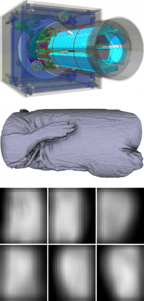
A plenoptic imaging system for preclinical in vivo imaging application has been developed (figure left). When imaging a three-dimensional (3D) object from six plenoptic camera positions, corresponding raw plenoptic data under bright-field illumination are shown. Acquisition setup and data used from a simulation study: Imaging system consisting of 6 plenoptic cameras with sensitive areas of 25mm× 50 mm, each, with nearly orthographic projections; there are (retractable) diffuse fiber line sources of detector length for object illumination located in the intermediary spaces between light detectors (top). The segmented volume of a reconstructed x-ray CT mouse data set is used as identified input (middle). Resulting raw plenoptic camera data as detected from 6 projection angles around 360° (bottom).
© dkfz.de
Computational mathematics
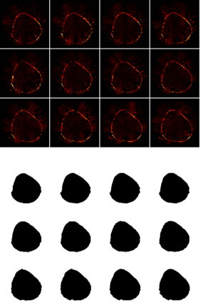
There are multiple imaging strategies involved in computational imaging. Here, surface reconstruction from multiview projection plenoptic image data is described (reference). The technique is adapted for in vivo small animal imaging, specifically imaging of nude mouse, and does not require an additional imaging step (e.g. be means of a secondary structural modality) or additional hardware (e.g. laser scanning approaches). Any potential point within the field-of-view (FOV) is evaluated by a proposed photo-consistency measure utilizing sensor image light information as provided by elemental images (EI's). As the superposition of adjacent EI's yields complementary information for any point within the FOV the three dimensional (3D) surface of the imaged object is estimated by a graph-cuts based method through global energy minimization. The proposed surface reconstruction is evaluated on simulated MLA-D data incorporating a reconstructed mouse data volume as acquired by X-ray CT. Compared to a previously presented back-projection based surface reconstruction method the proposed technique yields a significantly lower error rate. Moreover while the back-projection based method may not be able to resolve concave areas, the novel approach does. Our results further indicate that the proposed method achieves high accuracy at a low number of projections.