Research
- Research Topics
- Cell Biology and Tumor Biology
- Stem Cells and Cancer
- Inflammatory Stress in Stem Cells
- Experimental Hematology
- Molecular Embryology
- Signal Transduction and Growth Control
- Epigenetics
- Redox Regulation
- Vascular Oncology and Metastasis
- Clinical Neurobiology
- Molecular Neurogenetics
- Chaperones and Proteases
- Vascular Signaling and Cancer
- Molecular Neurobiology
- Mechanisms Regulating Gene Expression
- Molecular Biology of Centrosomes and Cilia
- Dermato-Oncology
- Pediatric Leukemia
- Tumour Metabolism and Microenvironment
- Personalized Medical Oncology
- Molecular Hematology - Oncology
- Cancer Progression and Metastasis
- Translational Surgical Oncology
- Neuronal Signaling and Morphogenesis
- Cell Signaling and Metabolism
- Cell Fate Engineering and Disease Modeling
- Cancer Drug Development
- Cell Morphogenesis and Signal Transduction
- Functional and Structural Genomics
- Molecular Genome Analysis
- Molecular Genetics
- Pediatric Neurooncology
- Cancer Genome Research
- Chromatin Networks
- Functional Genome Analysis
- Theoretical Systems Biology
- Neuroblastoma Genomics
- Signaling and Functional Genomics
- Signal Transduction in Cancer and Metabolism
- RNA Biology and Cancer
- Systems Biology of Signal Transduction
- Areas of Interest
- Advancement of clinical proteomics for systems medicine
- Bridging from the single cell to the cell population Epo-induced cellular responses and erythroleukemia
- Deciphering tumor microenvironment interactions determining lung cancer development
- Mechanisms controlling the compensation of liver injury and towards model-based biomarkers for early detection of liver cancer
- Application of dynamic pathway modelling for personalized medicine
- Group Members
- Publications
- Open Positions
- Funding
- Teaching
- Areas of Interest
- Molecular thoracic Oncology
- Proteomics of Stem Cells and Cancer
- Computational Genomics and System Genetics
- Applied Functional Genomics
- Applied Bioinformatics
- Translational Medical Oncology
- Metabolic crosstalk in cancer
- Pediatric Glioma Research
- Cancer Epigenomics
- Translational Pediatric Sarcoma Research
- Artificial Intelligence in Oncology
- Mechanisms of Genomic Variation and Data Science
- Neuropathology
- Pediatric Oncology
- Neurooncology
- Somatic Evolution and Early Detection
- Translational Control and Metabolism
- Soft-Tissue Sarcoma
- Precision Sarcoma Research
- Brain Mosaicism and Tumorigenesis
- Mechanisms of Genome Control
- Translational Gastrointestinal Oncology and Preclinical Models
- Translational Lymphoma Research
- Mechanisms of Leukemogenesis
- Genome Instability in Tumors
- Developmental Origins of Pediatric Cancer
- Brain Tumor Translational Targets
- Translational Functional Cancer Genomics
- Regulatory Genomics and Cancer Evolution
- SPRINT
- Cancer Risk Factors and Prevention
- Cancer Epidemiology
- Biostatistics
- Clinical Epidemiology and Aging Research
- Health Economics
- Physical Activity, Prevention and Cancer
- Preventive Oncology
- Personalized Early Detection of Prostate Cancer
- Digital Biomarkers for Oncology
- Genomic Epidemiology
- Cancer Survivorship
- Immunology and Cancer
- Cellular Immunology
- Molecular Oncology of Gastrointestinal Tumors
- Immunoproteomics
- T Cell Metabolism
- Personalized Immunotherapy
- mRNA Cancer Immunotherapies
- Translational Immunotherapy
- B Cell Immunology
- Immune Diversity
- Structural Biology of Infection and Immunity
- Applied Tumor Immunity
- Neuroimmunology and Brain Tumor Immunology
- Adaptive Immunity and Lymphoma
- Dermal Oncoimmunology
- Immune Regulation in Cancer
- Systems Immunology and Single Cell Biology
- GMP & T Cell Therapy
- Immune Monitoring
- News
- Imaging and Radiooncology
- Radiology
- Research
- Computational Radiology Research Group
- Contrast Agents In Radiology Research Group
- Neuro-Oncologic Imaging Research Group
- Radiological Early Response Assessment Of Modern Cancer Therapies
- Imaging In Monoclonal Plasma Cell Disorders
- 7 Tesla MRI - Novel Imaging Biomarkers
- Functional Imaging
- Visualization And Forensic Imaging
- PET/MRI
- Dual- and Multienergy CT
- Radiomics Research Group
- Prostate Research Group
- Breast Imaging Research Group
- Bone marrow
- Musculoskeletal Imaging
- Microstructural Imaging Research Group
- Staff
- Patients
- Research
- Medical Physics in Radiology
- X-Ray Imaging and Computed Tomography
- Federated Information Systems
- Translational Molecular Imaging
- Medical Physics in Radiation Oncology
- Biomedical Physics in Radiation Oncology
- Intelligent Medical Systems
- Medical Image Computing
- Radiooncology - Radiobiology
- Smart Technologies for Tumor Therapy
- Radiation Oncology
- Molecular Radiooncology
- Nuclear Medicine
- Translational Radiation Oncology
- Molecular Biology of Systemic Radiotherapy
- Interactive Machine Learning
- Multiparametric methods for early detection of prostate cancer
- Molecular Mechanisms of Head and Neck Tumors
- Radiology
- Infection, Inflammation and Cancer
- Tumor Virology
- Viral Transformation Mechanisms
- Pathogenesis of Virus-Associated Tumors
- Immunotherapy and Immunoprevention
- Applied Tumor Biology
- Virotherapy
- Virus-associated Carcinogenesis
- Chronic Inflammation and Cancer
- Microbiome and Cancer
- Cell Plasticity and Epigenetic Remodeling
- Experimental Hepatology, Inflammation and Cancer
- Infections and Cancer Epidemiology
- Tumorvirus-specific Vaccination Strategies
- Mammalian Cell Cycle Control Mechanisms
- Molecular Therapy of Virus-Associated Cancers
- DNA Vectors
- Episomal-Persistent DNA in Cancer- and Chronic Diseases
- Cell Biology and Tumor Biology
- Research Groups A-Z
- Junior Research Groups
- Core Facilities
- Center for Preclinical Research
- Chemical Biology Core Facility
- Electron Microscopy
- Flow Cytometry
- Genomics and Proteomics
- Information Technology
- Library
- Kataloge -- Catalogues
- Zeitschriften - Journals
- E-Books - ebooks
- Datenbanken - Databases
- Dokument-Lieferung - Document Delivery
- Publikationsdatenbank - Publication database
- DKFZ Archiv - DKFZ Archive
- Open Access
- Science 2.0
- Ansprechpartner - Contact
- More Information - Service
- Anschrift - Address
- Antiquariat - Second Hand
- Aufstellungssystematik - Shelf Classification
- Ausleihe - Circulation
- Benutzerhinweise - Library Use
- Beschaffungsvorschläge - Desiderata
- Fakten und Zahlen - Facts and Numbers
- Kooperationen, Konsortien - Cooperations, Consortia
- Kopieren, Scannen - Copying, Scans
- Kurse, Führungen - Courses, Introductions
- DKFZ-Intern - internal only
- DEAL-Info
- Light Microscopy
- Omics IT and Data Management Core Facility
- Small Animal Imaging
- Metabolomics Core Technology Platform
- Data Science @ DKFZ
- INFORM
- Baden-Württemberg Cancer Registry
- Cooperations & Networks
- National Cooperations
- International Cooperations
- Cooperational Research Program with Israel: DKFZ - MOST in Cancer Research
- Program
- Members of the Program Committee
- Call
- Publication Database
- German-Israeli Cancer Research Schools
- Archive
- Heidelberg - Israel, Science and Culture
- Symposium 40 Years of German-Israeli Cooperation
- 35th Anniversary Symposium
- 34th Meeting of the DKFZ-MOST Program
- 40th Anniversary Publication
- 30th Anniversary Publication
- 20th Anniversary Publication
- Flyer - The Cancer Cooperation Program
- List Publications 1976-2004
- Highlight-Projects
- Cooperational Research Program with Israel: DKFZ - MOST in Cancer Research
- Cooperations with industrial companies
- DKFZ PostDoc Network
- Cross Program Topic RNA@DKFZ
- Cross Program Topic Epigenetics@dkfz
- Cross Program Topic Single Cell Sequencing
- WHO Collaborating Centers
- DKFZ Site Dresden
- Health + Life Science Alliance Heidelberg Mannheim
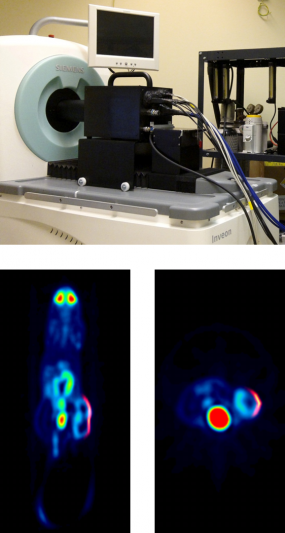
Synchromodal emission imaging
© dkfz.de
To enable what we call synchromodal optical imaging we invented the first plenoptic camera for diagnostic research that not only creates three-dimensional (illumination and fluence) projections of the imaged subject, but can also be integrated and used with PET and MRI (reference). The world's first simultaneously acquired [18F]FDG PET - pVEGF Luc BLI plenoptic image is shown in the left figure (top: experimental plenoptic system integrated into a Siemens Inveon PET for simultaneous data acquisition, bottom: fused PET and OI data (reference)).
A complete instrumentation and mathematical framework for preclinical plenoptic imaging (POI) is developed in which optical data is acquired by means of a microlens array (MLA) based light detector (MLA-D). The MLA-D has been developed to enable unique POI, especially in synchromodal operation with secondary imaging modalities such as PET or MRI. An MLA-D consists of a (large-area) photon sensor array, a matched MLA for field-of-view definition, and a septum mask of specific geometry made of anodized aluminum that is positioned between the sensor and the MLA to suppresses light cross-talk and to shield the sensor's radiofrequency interference signal (essential when used inside an MRI system). The software framework, while freely parameterizable for any MLA-D, is tailored towards a POI prototype system for preclinical synchromodal imaging application comprising a multitude of cylindrically assembled, gantry-mounted, simultaneously operating MLA-D's. When used in synchromodal operation, reconstructed tomographic volume data can be used for co-modal image fusion and also as a prior for estimating the imaged object's 3D surface by means of gradient vector flow. Superimposed planar or surface-aligned inverse mapping can be performed to estimate and to fuse the emission light map with the boundary of the imaged object. Triangulation and subsequent optical reconstruction can be performed to estimate the internal three-dimensional emission light distribution. The framework is susceptible to a number of variables controlling convergence and computational speed.