MSK Working Group
The joint working group of Musculoskeletal imaging at the Department of Radiology at the German Cancer Research Centre (DKFZ) and the Section of Musculoskeletal Radiology of the Department of Diagnostic and Interventional Radiology (DIR) at Heidelberg University Hospital is engaged in the implementation of innovative methods for functional multimodal and high-volume imaging of the musculoskeletal system in cooperation with the Department of Medical Physics in Radiology at the DKFZ.
The clinical and scientific focus of the MSK working group is the establishment of modern MRI and ultrasound techniques for cartilage, muscle, vascular and tumor imaging diagnostics. In addition to the collaboration on projects since 2012, a regular staff rotation between the Department of Radiology at the DKFZ and the Section of Musculoskeletal Radiology of DIR has been established in the curriculum.
Projects
High-field-MRI for the evaluation of joint cartilage vitality and integrity
Contact persons: Dr. med. Christoph Rehnitz, Dr. rer. nat. Armin M. Nagel
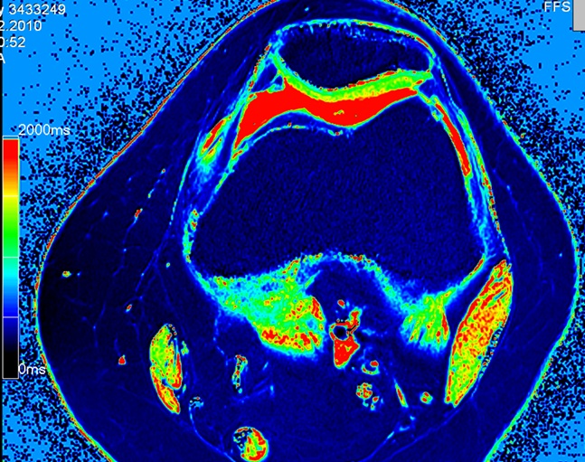
Image 1: dGEMRIC-sequence of the knee. Normal cartilage is depicted green. Areas of cartilage degeneration would appear blue.
In modern imaging of articular cartilage, in addition to the high-resolution morphological imaging, new developments arise that make it possible to visualize biochemical components of cartilage. These techniques include the so-called dGEMRIC technique and T2 mapping. Some of these biochemical components play a central role in the development of osteoarthritis or during cartilage healing following cartilage therapy. Therefore, the imaging morphology and quantification of cartilage components allows conclusions about the cartilage vitality (fig. 1) and integrity. The goal is to visualize early cartilage changes that are not yet visible by conventional MRI methods. Current main areas of application include: assessment of functional cartilage before surgery and follow-up examinations after cartilage therapy. Current projects investigate primarily the value of the 7-Tesla MRI.
Implementation of functional MRI-methods to evaluate normal and pathological skeletal muscle
Contact persons: Prof. Dr. med. M.-A. Weber, Dr. rer. nat. Armin M. Nagel
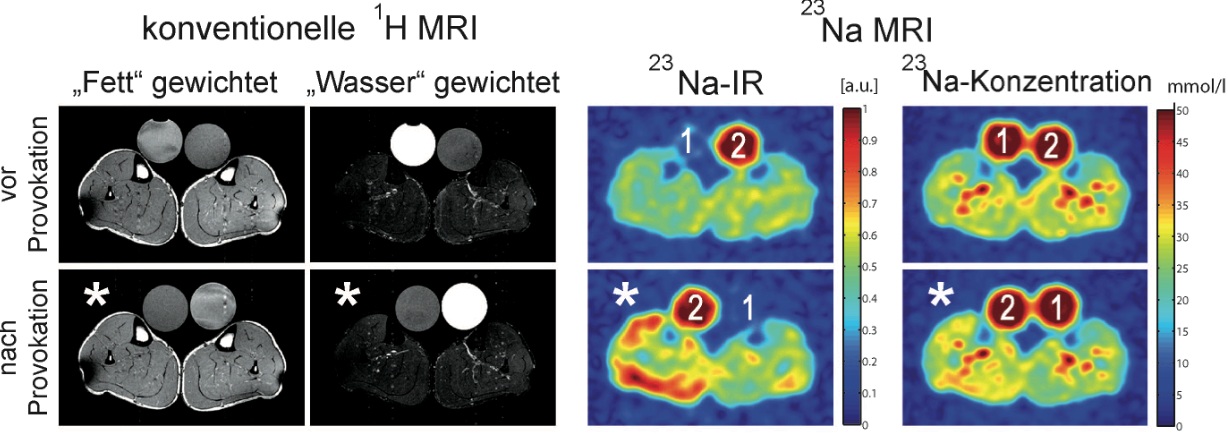
Image 2: Conventional 1H MRI (left) and 23Na MRI (right) of a patient with sodium-channel myopathy HyperPP. Following provocation by cooling of the right limb a signal increase in the 23Na-IR as a sign of a pathological intracellular Na + influx is shown.
With common radiological methods only morphological changes of the muscles can be visualised. New methods such as 23Natrium magnetic resonance imaging now provide information about pathophysiological changes. For example, the calf muscles of patients with paramyotonia or hyperkalemic periodic paralysis (HyperPP), both diseases caused by defective sodium channels of muscle resulting in intermittent paralysis, look completely normal on a conventional magnetic resonance image. Only the specific 23Natrium-MRI is able to visualize the pathologically increased sodium content of the muscle cells.
High-field-MRI in cartilage bone tumor diagnostics
Contact persons: Dr. rer. nat. Armin M. Nagel, Dr. med. Björn Jobke
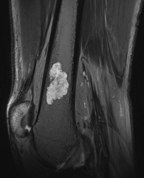
Image 3: Enchondroma of the distal Femur (7 Tesla; matrix 1024 x 512)
© Dr. med. Björn Jobke
The incidence of benign cartilaginous tumors (enchondroma) is about 5%, so that the incidental finding of this entity is fairly common (appr. weekly in our institute). In some cases the sole morphological differentiation of benign and less aggressive cartilage tumors (chondrosarcoma grade 1) is extremely difficult and even for histopathologists after biopsy a big challenge. Modern radiology can contribute information with high-resolution imaging, e.g. 7T high-field MRI, for a better characterization of the tumor growth behavior with respect to the surrounding tissue. The aim is to image tumor invasiveness on a microstructure level, similar to histology.

Image 4: Signal increase of an intra-osseous cartilage tumor (enchondroma) demonstrating increased sodium content in the cartilage matrix.
23Na is a component of glycoproteins, called proteoglycans, which play a crucial role in the formation of the cartilage matrix. 23Na can be imaged using high-field MRI and found in high concentration both in healthy cartilage, as well as in cartilage tumors, e.g. in enchondroma as demonstrated. This project deals with the analysis of 23Na concentration and their utility in the differential diagnosis and grading of cartilaginous tumors (Fig. 3 and Fig. 4) in collaboration with the Department "Medical Physics in Radiology" (E020).
Imaging in monoclonal plasma cell disease
Contact persons: Prof. Dr. med. Stefan Delorme, Dr. med. Jennifer Mosebach
Fusions-Ultrasound (eSie) in the diagnostics of peripheral soft tissue sarcoma
Contact persons: Dr. med. Björn Jobke, Dr. med. Jessica Jesser, Prof. Dr. med. M.-A. Weber
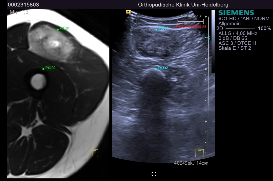
Image 5: Fusion-based sonography. Left: MRI soft-tissue tumor of the thigh; Right: Parallel ultrasound
A new ultrasound system (Acuson S3000 eSie, Siemens) is capable of displaying ultrasound images simultaneously to cross-sectional images. It enables an overlay or parallel visualization of real-time ultrasound images with existing MRI and CT scans of the same anatomical region. This so-called fusion imaging allows for a quick anatomical orientation and provides more imaging details for the interpretation of complex pathologies or in order to perform interventions such as tumor biopsies. We currently carry out a feasibility study to assess the value in the routine diagnostics of peripheral soft tissue tumors. Both technical aspects and the learning curve for this new technology will be investigated. In partnership with the manufacturer and developer it is our goal to improve the system in order to use it as primary diagnostic tool and during guidance of ultrasound-assisted biopsies. Especially the tissue biopsy sampling of heterogeneous soft tissue tumors is extremely important for the diagnosis and grading. Here, a targeted approach to a representative tissue biopsy with MRI information of soft tissue composition, using fusion ultrasound, may be of great diagnostic value.
Publications
3 Tesla sodium inversion recovery magnetic resonance imaging allows for improved visualization of intracellular sodium content changes in muscular channelopathies.
Nagel AM, Amarteifio E, Lehmann-Horn F, Jurkat-Rott K, Semmler W, Schad LR, Weber MA
Invest Radiol. 2011 Dec;46(12):759-66
Hyperkalemic periodic paralysis and permanent weakness: 3-T MR imaging depicts intracellular 23Na overload initial results.
Amarteifio E, Nagel AM, Weber MA, Jurkat-Rott K, Lehmann-Horn F
Radiology. 2012 Jul;264(1):154-63
Knochentumoren in 'Radiologische Diagnostik der Knochen und Gelenke' (Bohndorf, Jobke et al., 3. Auflage, Thieme Verlag)