Group E0406: Novel Detection Techniques for Ion Beams
Group Leader: Dr. Mária Martiíková

Ion beam radiotherapy & its challenges
Ion beam radiotherapy is a highly precise treatment modality for tumors close to critical organs like brainstem or eye nerves. Unlike in conventional radiotherapy with photons, in ion radiotherapy even minor geometrical changes in the patient's anatomy (e.g. tissue swelling, different kinds of movement or incorrect patient positioning) can lead to severe changes of the radiation dose distribution in the patient. Consequently, quantitative imaging of the internal patient structures and the radiation field inside of the patient during therapy are crucial for the success of ion-beam treatments.
The goal of our group is to decrease the uncertainties of the dose distribution in the patient, in order to exploit the full therapeutic potential of ion beam radiotherapy. In our research we closely collaborate with the Heidelberg Ion Beam Therapy Center (HIT).
Timepix detectors:
In all our projects Timepix detectors are used for the experimental measurements. The generations of Timepix radiation detector technology were developed at CERN. These finely pixelated semiconductor detectors enable noise-free detection of individual ionizing particles and their properties, as well as ion tracking.
Current research projects
➥In-ViMo project: Non-contact assessment of the dose distribution in the patient
Secondary radiation is used in this project for in-vivo monitoring of the dose distribution in the patient. As secondary radiation is escaping the patient instantaneously during the irradiation as a by-product, no additional dose to the patient is required for this kind of imaging.
While a wide spectrum of radiation is created in the patient, we focus on measuring the tracks of charged nuclear fragments. By analysing their distribution we draw conclusions about internal changes in the patient's tissue along the beam path (e.g. filling of the nose cavity).
Following year-long research on patient models, we started a clinical study at the Heidelberg Ion Beam Therapy Center.
Funding: National Center for Tumor Diseases (NCT), Funding Program "Proof-of-Concept Clinical Trials", Project: In-vivo monitoring of carbon ion radiotherapy delivery (InViMo)
Related publications:
1. L. Ghesquière-Diérickx, R. Félix-Bautista, A. Schlechter, L. Kelleter, M. Reimold, G. Echner, P. Soukup, O. Jäkel, T. Gehrke and M. Martiíková. Detecting perturbations of a radiation field inside a head-sized phantom exposed to therapeutic carbon-ion beams through charged-fragment tracking. Medical Physics 29 (2022) 1776-1792 Editor's Choice
2. L. Ghesquière-Diérickx, A. Schlechter, R. Félix-Bautista, T. Gehrke, G. Echner, L. Kelleter and M. Martiíková. Investigation of Suitable Detection Angles for Carbon-Ion Radiotherapy Monitoring in Depth by Means of Secondary-Ion Tracking. Frontiers in Oncology 11 (2021) 780221
3. R. Félix‐Bautista, L. Ghesquière‐Diérickx, L. Marek, C. Granja, P. Soukup, D. Turecek, L. Kelleter, S. Brons, M. Ellerbrock, O. Jäkel, T. Gehrke and M. Martiíková. Quality assurance method for monitoring of lateral pencil beam positions in scanned carbon‐ion radiotherapy using tracking of secondary ions. Medical Physics 8 (2021) 4411-4424
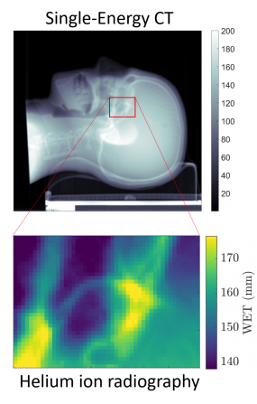
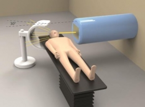
➥He-ion radiography: Imaging of the patient with helium ion beams
To precisely identify possible changes within the patient directly before the treatment, when the patient is already positioned on the couch, we develop an imaging method with helium ions instead of conventionally used photons. These ions must have sufficient energy to cross the entire patient, since the residual energy of the ions leaving the patient is needed. Bulky calorimeters are used to measure it. Our approach is to use thin (< 1 mm) Timepix detectors, in combination with an adjustable energy of the incoming helium-ion beam. This unique approach we named "energy painting".
Funding: German Research Society (DFG), Project:Ion radiography for motion detection (Grant ID: MA 4437/3-1) and Project: Ion radiography with adaptive energy choice for a precise radiotherapy
Related publications:
1. C. Knobloch, M. Metzner, F. Kehrein, C. Schömers, S. Scheloske, S. Brons, R. Hermann, A. Peters, O. Jäkel, M. Martiíková and T. Gehrke. Experimental helium-beam radiography with a high-energy beam: Water-equivalent thickness calibration and first image-quality results. Medical Physics 49 (2022) 5347-5362 Editor's Choice
2. C. Amato, M. Martiíková, and T. Gehrke. A technique for spatial resolution improvement in helium‐beam radiography. Medical Physics 5 (2020) 2212-2221
3. M. Martiíková, T. Gehrke, S. Berke, G.Aricò and O. Jäkel. Helium ion beam imaging for image guided ion radiotherapy. Radiation Oncology 13(1) (2018) 109
➥Ion sprectroscopy: Characterization of mixed radiation fields for determination of biological effects
In clinical practice the proper radiation dose administered to a patient is examined in detail on multiple steps of the therapeutic chain. The main physical parameter characterizing the radiation quality, the so called "linear energy transfer", is crucial for calculation of biological effects of the radiation. Currently, it is estimated only by means of simulations due to the lack of suitable measurement methods.
We are developing a robust method to measure several radiation quality parameters simultaneously. We use the capability of the Timepix detectors to measure the path of individual ions, their type and energy which each ion deposits in the detector. The patient-to-patient assessment of the radiation quality will enable in the future to tailor the radiation effect more precisely to the irradiated tissue.
This research is closely connected to dosimetry in outer space, in particular the determination of the effective biological dose to which astronauts are exposed to.
Funding: German Cancer Aid (Deutsche Krebshilfe), Project: Development of a method for high resolution verification of the beam quality in ion beam radiotherapy (Grant ID: 70114370 ) and Project: Ion spectroscopy for improvement of the physical beam model for therapy planning in ion beam therapy (Grant ID: 110296)
Related publications:
1. G. Arico, T. Gehrke, R. Gallas, A. Mairani, O. Jäkel and M. Martisikova. Investigation of single carbon ion fragmentation in water and PMMA for hadron therapy. Physics in Medicine and Biology 64 (2019) 055018
2. C. Granja, J. Jakubek, M. Martisikova, S. Kodaira, S. Polansky, P. Krist, V. Zach and T. Matlocha. Dynamic range and resolving power of the Timepix detector to heavy charged particles. Journal of Instrumentation 13 (2018) C11003
3. T. Gehrke, L. Burigo, G. Arico, S. Berke, J. Jakubek, D. Turecek, T. Tessonnier, A. Mairani and M. Martiíková. Energy deposition measurements of single 1H, 4He and 12C ions of therapeutic energies in a silicon pixel detector. Journal of Instrumentation 12 (2017) P04025
Further Information
Main cooperation partners:

We thank our funding bodies:
- National Center for Tumor Diseases (Nationale Centrum für Tumorerkrankungennal - NCT)
- German Science Society (Deutsche Forschungsgemeinschaft - DFG)
- German Cancer Aid (Deutsche Krebshilfe)
- Excellence Initiative of the Heidelberg University
- Heidelberg University Hospital (Universitätsklinikum Heidelberg - UKHD)
- German Cancer Research Center (Deutsches Krebsforschungszentrum - DKFZ)
- German Academic Exchange Service (Deutsches Akademisches Austauschdienst - DAAD)