ARTEMIS Adaptive RadioThErapy with MR guided Ion beamS
ARTEMIS Adaptive RadioThErapy with MR guided Ion beamS
© dkfz.de
The ARTEMIS project focuses on the development of image-guided adaptive radiotherapy for ion beams and its clinical introduction. In addition to data from X-ray computed tomography (CT), data from magnetic resonance imaging (MRI) will be made available for the therapy and used during the course of therapy. The particular advantage of this approach is that, due to the improved image information, a more precise and gentler therapy is possible. A combination with MRI should help to better detect changes in the tumor during therapy and consequently further optimize irradiation without increasing the dose.
The ARTEMIS project is a collaboration between the Department of Radiation Oncology and Radiotherapy (Head: Prof. Dr. Dr. Jürgen Debus) at Heidelberg University Hospital (UKHD) and the Division of Medical Physics in Radiation Oncology (Head: Prof. Dr. Oliver Jäkel) at the German Cancer Research Center (DKFZ). The funding from the Federal Ministry of Education and Research (FMER, grant number: 13GW0436B) ran at the DKFZ from 01.08.2019 to 31.08.2023. The funding at the UKHD (grant number 13GW0436A) ends in 2024.
Leaders

Prof. Oliver Jäkel, PhD and Prof. Jürgen Debus, MD, PhD
© DKFZ & UKHD
Professor Jürgen Debus, MD, PhD
Medical Director of the Heidelberg Ion-Beam Therapy Center (HIT) and of the Radiation Oncology and Radiation Therapy Clinic at the Heidelberg University Hospital
Professor Oliver Jäkel, PhD
Head of the Department of Medical Physics in Radiation Oncology, DKFZ, Heidelberg
Head of Medical Physics of the Heidelberg Ion-Beam Therapy Center (HIT)
Sub-Projects at DKFZ and UKHD
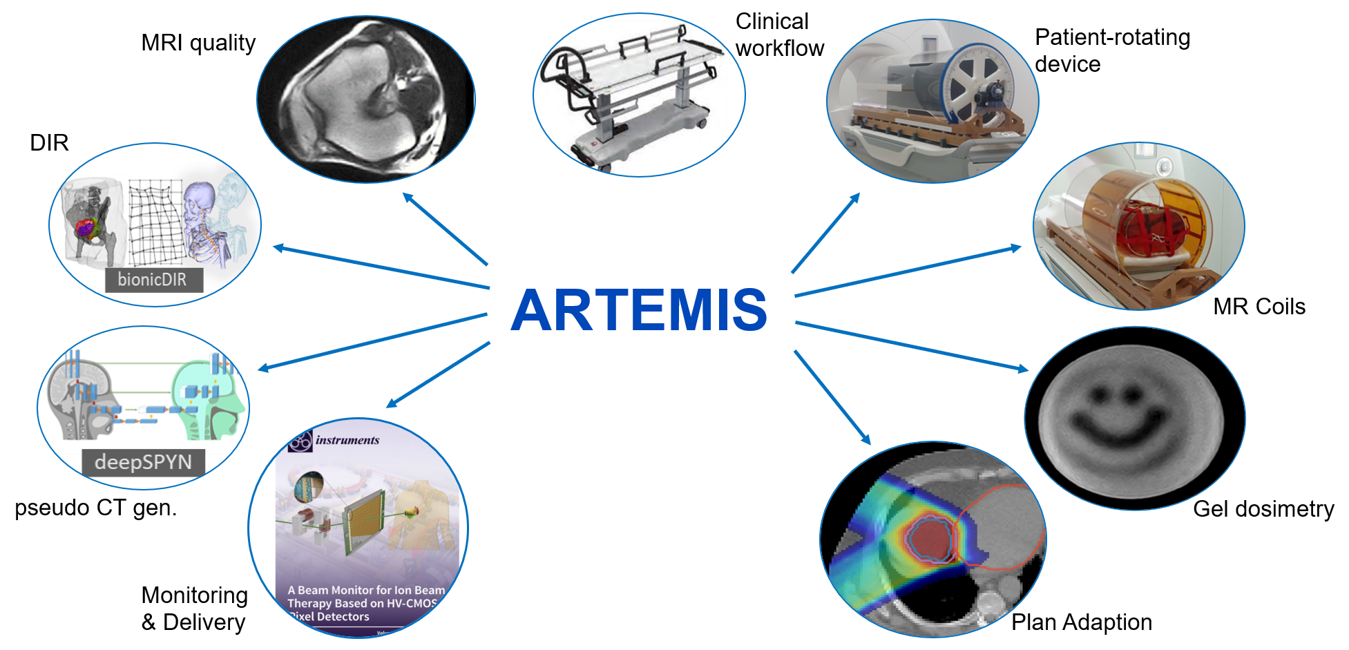
The overall goal of the project is to develop a functional demonstrator for MR-guided ion irradiation at the Heidelberg Ion Beam Therapy Center (HIT). The project comprised the following 10 sub-projects (SP):
- Development of an MRI system at HIT (UKHD)
- Adaptation of the beam application system and the monitoring system (BAMS) (UKHD)
- Development of a patient positioning system (UKHD)
- Development of a rotatable patient positioning system with MR coil adapters (DKFZ)
- Development and testing of software for correction and conversion of MR data in pseudo-CT for BPL of ion beams (DKFZ)
- Optimization and quality assurance of imaging for MRgIT (DKFZ)
- Adaptation of the existing irradiation planning to the field strength and geometry of the magnet and online optimization of the irradiation field (UKHD)
- Development of the imaging coils for use in radiation planning and irradiation (UKHD)
- Accompanying the setup through quality management and quality assurance (UKHD)
- Clinical specification and implementation of clinical workflows (UKHD)
Sub-Projects at DKFZ
Subprojects 4-6 at DKFZ were successfully completed on August 31, 2023. These included the development of a novel, rotatable patient positioning system (SP 4). As main result, a rotatable patient capsule for clinical studies was made available at UKHD, which has already been used for initial volunteer studies and is now to be used in clinical studies for evaluation (see here).
In addition, algorithms for MRI-based therapy planning and methods for optimizing MRI image data were developed (SP 5). Here, software-based solutions for data analysis, image registration and generation of so-called pseudo-CTs were implemented in order to be able to carry out a movement analysis with the MR data obtained, which in turn serve as a starting point for adapting the therapy plan in the course of therapy (see here).
Another component was the optimization of quality assurance for the MR-guided therapy process (SP 6). This involved optimizing the MR image sequences for a study protocol that acquired patient data for the aforementioned data analysis (SP 5). In addition to optimizing the MRI sequences for applications in particle therapy and developing a quality assurance protocol, the long-term stability of the MR scanners used and the automation of quality assurance were investigated. Another result is the development of an automated tool for MR sequence optimization (see here).
Furthermore, the development of imaging coils for use in radiation planning and radiation (SP 8) was carried out in close cooperation between the UKHD and the Division of Medical Physics in Radiology at the DKFZ.
Publications
Journal Articles
Bauer C et al. 2023 Biofidelic image registration for head and neck region utilizing an in-silico articulated skeleton as a transformation model. Physics in Medicine & Biology, Volume 68, Number 9, DOI: 10.1088/1361-6560/acc7f1
Dorsch S et al. 2023 Quality assurance and temporal stability of a 1.5 T MRI scanner for MR-guided Photon and Particle Therapy, Z Med Phys 2023, DOI: 10.1016/j.zemedi.2023.04.004
Elter A et al. 2021 Development of phantom materials with independently adjustable CT- and MR-contrast at 0.35, 1.5 and 3 T, Phys Med Biol. 2021, DOI: 10.1088/1361-6560/abd4b9
Fahad H et al. 2023 Multi-parametric optimization of MR-imaging sequences for MR-guided radiotherapy, phiRo 2023, https://doi.org/10.1016/j.phro.2023.100497
Marot M et al. 2023 Proton beam dosimetry in the presence of magnetic fields using Farmer-type ionization chambers of different radii, Medical Physics 2023, DOI: 10.1002/mp.16368
Marot M et al. 2023 Monte Carlo simulation for proton dosimetry in magnetic fields: Fano test and magnetic field correction factors kB for Farmer-type ionization chambers. Phys Med Biol. 2023, doi: 10.1088/1361-6560/acefa1
Putz F et al. 2024 Quality requirements for MRI simulation in cranial stereotactic radiotherapy: a guideline from the German Taskforce Imaging in Stereotactic Radiotherapy. Strahlentherapie und Onkologie 200, 1-18, 2024; https://link.springer.com/article/10.1007/s00066-023-02183-6
Walter A et al. 2024 Segmentation of seventy-one anatomical structures necessary for the evaluation of guideline-conform clinical target volumes in head and neck cancers. Cancers 2024, 16(2), 41, https://doi.org/10.20944/preprints202312.1562.v1
Weidner A et al. 2022 An abdominal phantom with anthropomorphic organ motion and multimodal imaging contrast for MR-guided radiotherapy. Phys Med Biol. 2022, doi: 10.1088/1361-6560/ac4ef8
Yawson A et al. 2024 Essential parameters needed for a U-Net-based segmentation of individual bones on planning CT images in the head & neck region using limited datasets for radiotherapy application. Phys Med Biol. 2024, https://doi.org/10.1088/1361-6560/ad1996
Conference Contributions
Bauer C et al. Biomechanics in non-rigid image registration for adaptive radiotherapy of head and neck cancer: Articulated skeleton as an accurate transformation model 9th World Congress of Biomechanics, Taipeh, Taiwan, 2022 talk
Bauer C, et al. Synthetische Bilddaten Augmentierung für DL-basierte Segmentierung mit einem biomechanischen, kinematischen Patientenmodell. Proceedings of the 53rd Annual Scientific Meeting DGMP, Aachen 2022
Bauer C et al. CT-MR-Deformable Image Registration for MR-Guided Radiotherapy using a Biomechanical Skeleton Model 9th MR in RT Symposium, Los Angeles, United Stated of America, 2023 talk
Dietrich KA, Klüter S, Knowles BR, Debus J, Ladd ME, & Platt T. Comparison of birdcage resonator designs for clinical MR-guided radiotherapy. Proc Intl Soc Mag Reson. Med. 2021; 29: 4013.
Dietrich KA, Klüter S, Debus J, Dinkel F, Echner G, Ladd ME, & Platt T. Construction of a Tx/Rx body coil on a rotatable patient capsule for MR-guided particle therapy. Proc Intl Soc Mag Reson Med. 2022; 30: 1094.
Dietrich KA, Klüter S, Debus J, Ladd ME, & Platt T. Performance of a rotatable Tx/Rx body coil for MR-guided particle therapy. Proc Intl Soc Mag Reson Med. 2023; 31: 4385.
Dietrich KA, Orzada S, Fiedler TM, Dinkel F, Echner G, Klüter S, Debus J, Ladd ME, & Platt T. A radiation-transparent, rotatable receive-only extremity RF coil for flexible MR-guided particle therapy at 0.25 T. Proc Intl Soc Mag Reson Med. 2024; 32 (accepted).
Dorsch S: MRT-QA für die Strahlentherapie. DGMP 2020
Dorsch S et al. Measurement of isocenter accuracy and image distortions in MRgRT. MRinRT 2021
Dorsch S et al. Charakterisierung der MR-Bildqualität für gleichzeitige MR-Aufnahme und Strahlscanning in der Partikeltherapie. DGMP 2023
Dorsch S et al. Characterization of MR image quality for simultaneous ion beam scanning in particle therapy. MRinRT Symposium 2023
Echner G et al. 2023 Development and test of a patient immobilization and rotation device for imaging and radiotherapy Poster presentation ESTRO Congress 2023, Wien
Fahad H et al. Multi-parametric optimization of MR-imaging sequences for MR-guided radiotherapy. ESTRO 2022
Fahad H et al. Multi-parametric optimization of MR-imaging sequences for MR-guided radiotherapy. ESMRMB 2023
Giske K et al. Towards biomechanical thorax model for lung radiotherapy: Model-basedregistration with kinematic articulated skeleton. 9th World Congress of Biomechanics, Taipeh, Taiwan, 2022 invited talk
Paul K., Dorsch S., Naumann J., Hansmann T., Haberer T., Debus J., Klüter S.: Towards MR-guided article radiotherapy: Compatibility of an open MR scanner with an ion beamline. Proceedings of the ESTRO Conference, 6.-10. May 2022, Copenhagen, Danmark, OC-0778
Walter A, et al. The impact of additional MRI scans on training of supervised deep learning methods for automatic CTV delineation in head and neck cancer 9th MR in RT Symposium, Los Angeles, United Stated of America, 2023
Weidner A., Steng C., Dinkel F., Dorsch S., Runz A., Echner G.: Development of an anthropomorphic real-time breathing phantom for image-guided radiotherapy. Tagung der Deutschen Gesellschaft für Medizinische Physik, 27.-30.09.2023, Wien p.V36
Yawson A et al. U-net-Based Segmentation for Individual Bones in radiation therapy planning CT scans for Head & Neck region. Proceedings of the 26th Annual Conference, MIUA 2022, Cambridge, UK, 2022 poster presentation
Yawson A et al. Pseudo-SPR Map Generation from MRI Using U-Net Architecture for Ion Beam Therapy Application. In Medical Image Understanding and Analysis - 27th Annual Conference, MIUA 2023, Aberdeen, UK, July 1921, 2023, Proceedings MIUA 2023: 257-267 poster presentation
PhD Theses:
Bauer C. Applications of a Biomechanical Patient Model for Adaptive Radiation Therapy. Dissertationsschrift 2023. Fakultät für Physik und Astronomie, Universität Heidelberg
Elter A. Development of end-to-end tests for online adaptive magnetic resonanceguided radiotherapy, Dissertationsschrift 2022, Fakultät für Physik und Astronomie, Universität Heidelberg
Funding
The project is funded by the Federal Ministry of Education and Research (FMER, grant numbers: 13GW0436A (UKHD); 13GW0436B (DKFZ)).
